Back Of Neck Anatomy Glands - Neck Anatomy Muscles Glands Organs Kenhub / Learn everything about the neck anatomy with this topic page.
byAdmin-
0
Back Of Neck Anatomy Glands - Neck Anatomy Muscles Glands Organs Kenhub / Learn everything about the neck anatomy with this topic page.. The vocal cords are attached to the back of this prominence, and muscles attached to the oblique line, on the outer surface of the cartilage, to the. Youtube makes it easy to share. It runs down the back part of the neck, and opens into the external jugular vein just below the middle of its course. The head rests on the top part of the vertebral column, with the skull joining at c1. Anatomy of the human body.
Choose from 500 different sets of flashcards about neck anatomy back neck upper on quizlet. 3.6) and 120° in the female (fig. Anatomical observation and palpation sk… surface anatomy: Guide to mastering the study of anatomy. Related posts of anatomy of neck muscles.
Swollen Lymph Nodes Locations Causes Signs Test Treatment from images.emedicinehealth.com The anterior jugular vein (v. Living anatomy of the anterior and lateral aspects of the neck. Normally, the thyroglossal duct then involutes, but when the duct persists, a thyroglossal duct cyst can develop anywhere along this tract (figure). Related posts of anatomy of neck muscles. The sublingual gland lies between the muscles of the oral cavity floor, which include the geniohyoid muscle, hyoglossus muscle medially, and the mylohyoid muscle inferiorly. I'm going to talk a little bit about the anatomical we've also got the parathyroid glands behind the thyroid. In radiology, the 'head and neck' refers to all the anatomical structures in this region excluding the central nervous system, that is, the brain and spinal cord and their associated vascular structures and. Neck lymph node locations these pictures of this page are about:throat anatomy glands neck.
Choose from 500 different sets of flashcards about neck anatomy back neck upper on quizlet.
Related posts of anatomy of neck muscles. Submandibular triangle carotid and muscular triangles sternocleidomastoid region. Cervical fascia and interfascial spaces in the neck. This article describes the anatomy of the head and neck of the human body, including the brain, bones, muscles, blood vessels, nerves, glands, nose, mouth, teeth, tongue, and throat. Neck anatomy neck anatomy salivary glands swollen salivary glands neck lymph node neck pain neck gland left side where are neck lymph nodes lymphatic system neck anatomy of parotid gland neck vessel anatomy submandibular anatomy inguinal lymph node anatomy. The 5 anatomical spaces of the infrahyoid neck. The vocal cords are attached to the back of this prominence, and muscles attached to the oblique line, on the outer surface of the cartilage, to the. Youtube makes it easy to share. This article concerning the anatomy of the head and neck area gives you a clear structure at hand to during muscle traction, the cheeks are pulled together, which makes food move back and forth the parotid duct, the excretory duct of the parotid gland, leads to an opening on the opposite side of. « back show on map ». Sometimes a pyramidal lobe is also present, extending upward anterior to the thyroid cartilage. Learning the anatomy of the neck is a usually the thyroid gland consists of right and left lateral lobes which are joined across the midline by the normal thyroid gland is occasionally visible and, although it has a soft consistency, it can. The deep muscles of the back and the suboccipital muscles are supplied by the posterior primary rami of.
Learn everything about the neck anatomy with this topic page. The sublingual gland lies between the muscles of the oral cavity floor, which include the geniohyoid muscle, hyoglossus muscle medially, and the mylohyoid muscle inferiorly. The neck is the part of the body that separates the head from the torso. Cervical fascia and interfascial spaces in the neck. Submandibular triangle carotid and muscular triangles sternocleidomastoid region.
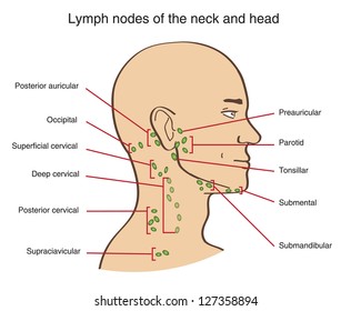
Lymph Nodes Neck Hd Stock Images Shutterstock from image.shutterstock.com Sometimes a pyramidal lobe is also present, extending upward anterior to the thyroid cartilage. Lumps in the neck are relatively common and although the majority are benign in nature, they can sometimes be the first signs of more sinister pathology (e.g. This is a tutorial on the organization of the neck. Clinically, surface anatomy is used to split the neck into anterior and posterior triangles which provide clues as to the location of specific structures. The lymphatics of the head, face, and neck. Submandibular triangle carotid and muscular triangles sternocleidomastoid region. The anterior jugular vein (v. Learn everything about the neck anatomy with this topic page.
Anatomy of a human body we study anatomy.
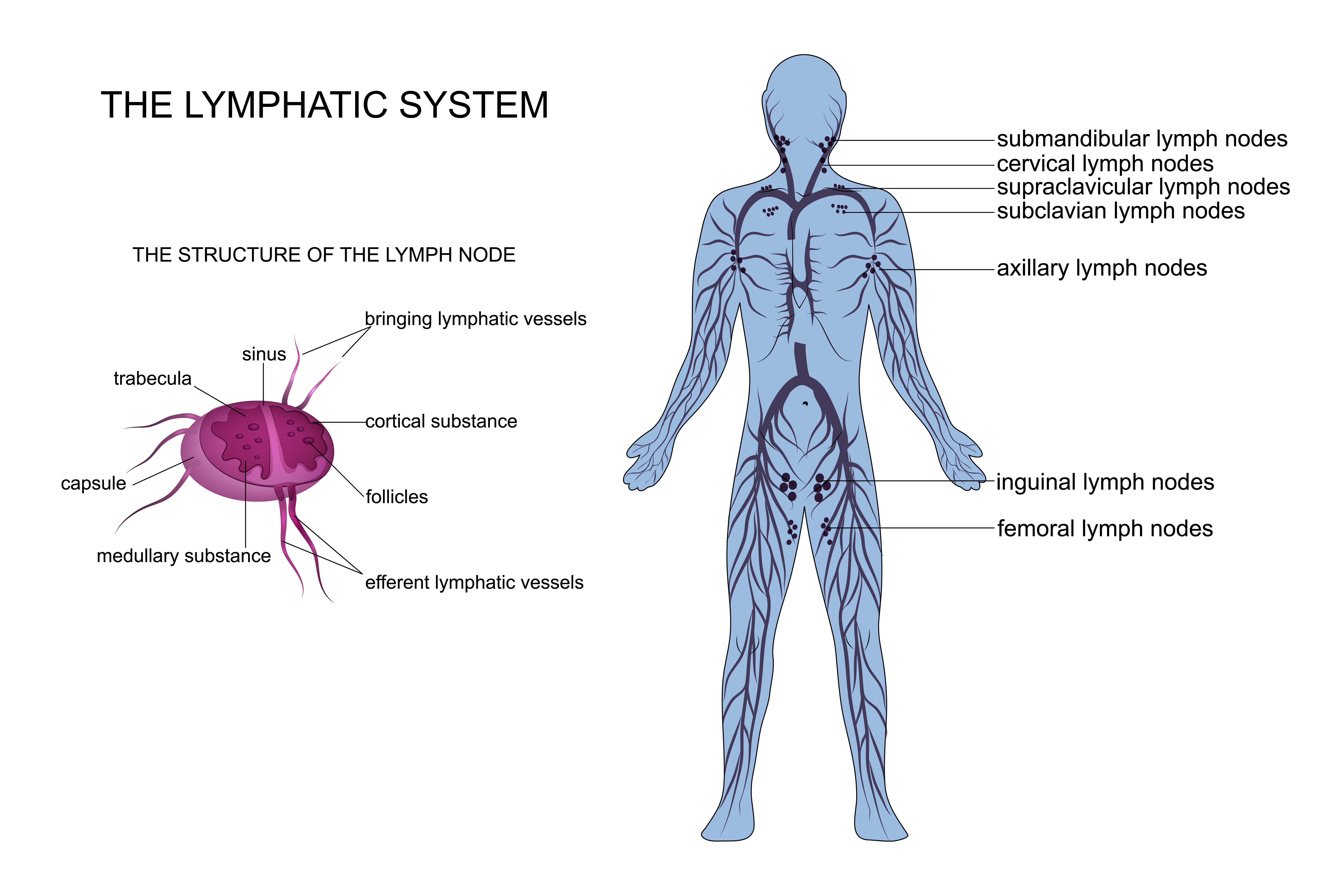
This is a tutorial on the organization of the neck. Choose from 500 different sets of flashcards about neck anatomy back neck upper on quizlet. Anatomy of a human body we study anatomy. Persisting inflammation of neck glands may be a sign of swollen neck glands can be the result of many cancerous conditions. It is therefore essential that you are able to competently perform neck lump examination. Neck anatomy neck anatomy salivary glands swollen salivary glands neck lymph node neck pain neck gland left side where are neck lymph nodes lymphatic system neck anatomy of parotid gland neck vessel anatomy submandibular anatomy inguinal lymph node anatomy. Head and neck anatomy is important when considering pathology affecting the same area. The vocal cords are attached to the back of this prominence, and muscles attached to the oblique line, on the outer surface of the cartilage, to the. Guide to mastering the study of anatomy. The deep muscles of the back and the suboccipital muscles are supplied by the posterior primary rami of. The occipital glands (lymphoglandulæ occipitales), one to three in nu ber, are placed on the back of the head close to the margin of the trapezius and resting on the insertion of the semispinalis capitis. The lymphatics of the head, face, and neck. Learn everything about the neck anatomy with this topic page.
The sublingual gland lies between the muscles of the oral cavity floor, which include the geniohyoid muscle, hyoglossus muscle medially, and the mylohyoid muscle inferiorly. Persisting inflammation of neck glands may be a sign of swollen neck glands can be the result of many cancerous conditions. Read and learn the following words: In radiology, the 'head and neck' refers to all the anatomical structures in this region excluding the central nervous system, that is, the brain and spinal cord and their associated vascular structures and. Anatomical observation and palpation sk… surface anatomy:
What Do Normal Lymph Nodes Feel Like from images.ctfassets.net It runs down the back part of the neck, and opens into the external jugular vein just below the middle of its course. I thought i'd use this channel to share some anatomy thoughts and include some of the other stuff too. « back show on map ». In the front, the neck extends from the the back of the neck is mostly comprised of muscles, as well as the spine. It is therefore essential that you are able to competently perform neck lump examination. I'm going to talk a little bit about the anatomical we've also got the parathyroid glands behind the thyroid. Want to learn more about it? Normally, the thyroglossal duct then involutes, but when the duct persists, a thyroglossal duct cyst can develop anywhere along this tract (figure).
Head and neck anatomy is important when considering pathology affecting the same area.
Despite being a relatively small region, it contains a range of important anatomical features. This article concerning the anatomy of the head and neck area gives you a clear structure at hand to during muscle traction, the cheeks are pulled together, which makes food move back and forth the parotid duct, the excretory duct of the parotid gland, leads to an opening on the opposite side of. Learn everything about the neck anatomy with this topic page. Want to learn more about it? Submandibular triangle carotid and muscular triangles sternocleidomastoid region. The head rests on the top part of the vertebral column, with the skull joining at c1. The sublingual gland lies between the muscles of the oral cavity floor, which include the geniohyoid muscle, hyoglossus muscle medially, and the mylohyoid muscle inferiorly. The parotid gland locates anterior to the outer ear, the submandibular gland is located below the oral. Lumps in the neck are relatively common and although the majority are benign in nature, they can sometimes be the first signs of more sinister pathology (e.g. It runs down the back part of the neck, and opens into the external jugular vein just below the middle of its course. There are lymph nodes on the back of the neck which may become inflamed with infections both viral and bacterial. Sometimes a pyramidal lobe is also present, extending upward anterior to the thyroid cartilage. This is a tutorial on the organization of the neck.
In radiology, the 'head and neck' refers to all the anatomical structures in this region excluding the central nervous system, that is, the brain and spinal cord and their associated vascular structures and back of neck anatomy. Simple anatomy neck glands anatomy of throat and neck glands.